The structure of the alimentary canal follows a consistent pattern from the level of oesophagus onwards
Modification from the general plan is due to the special function associated with those organs
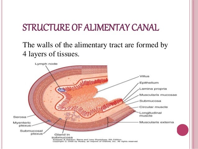
The wall of the alimentary tract is formed by 4 layers in sequence from outside as follows
Serosa/ Adventita
Muscularis Externa
Sub Mucosa
Mucosa
Major variation from the basic plan occurs generally in the mucosa
Serosa
Outermost layer generally has two layers outer peritoneum which is made up of simple squamous epithelium and fibrous connective tissue
Serosa forms the mesenteries which suspend and support the organs from the body wall
Muscle layer/ Muscularis Externa
This layer is composed of an outer longitudinal and an inner circular layer of smooth muscles between these two layers is an autonomic nerve plexus which is named as Auberch’s plexus which contains both sympathetic and parasympathetic nerves
Smooth muscles are innervated by this nerves
Coordinated and alternative contraction of longitudinal muscle fibre and circular muscle fibre occurs in Rhythm resulting the food to more along the alimentary canal. This type of contraction is called peristalsis
Circular layer of muscle becomes thicken at specific places to form spinsters
Parasympathetic impulses stimulate the movement of gut wall and make the spinsters to open while sympathetic impulses inhibit the peristaltic movement and cause the spinsters to close
Submucosa
Loose connective tissue containing large amount of collagen and elastin fibres, large blood vessels, lymph vessels.
Autonomic nerves which are passed to and from the mucosa between the circular muscle layer and submucosa is the submucosal meissner’s plexus which also contains both sympathetic and parasympathetic nerves
Mucosa
This consists of three layers of tissues
Muscularis mucosa
Lamina propria
Epithelial lining/ mucus membrane
Muscularis mucosa
This is thin layer of smooth muscle it is helped to bring about local movement of the mucosa independent from the rest of the parts of the wall thus bringing mucous membrane in close contact with the food
Lamina propria
It is made up of areolar tissue it has lymphoid tissue to product from invading microorganisms
Mucous membrane
This is the innermost layer
Has three main functions
protection
secretion
absorption
Found in parts of the tract which are subjected to wear and tear or mechanical injury
This layer consists of non-keratinized stratified squamous epithelium with mucus secreting glands in areas where the food is already soft and moist and where secretions of digestive tissues and absorption
Mucous membrane consists of simple columnar epithelial cells interspersed with mucus secreting Goblet cells